What is an Ultrasound?
The term “ultrasound” refers to sound with a frequency that humans cannot hear.
Ultrasound is a sound that travels through soft tissue and fluids, but it bounces back off denser surfaces and creates an image from the inside of your body. It’s also known as sonography and is a painless procedure.
Ultrasound uses no radiation and for this reason, it’s the preferred method for viewing a developing fetus during pregnancy.
Why an ultrasound is performed?
An ultrasound allows your doctor to see problems with organs, vessels, joints, tendons, muscles and other tissues without needing to make an incision.
An ultrasound can provide a view of the:
- Liver
- Gall Bladder
- Pancreas
- Spleen
- Kidneys
- Urinary Bladder
- Prostate
- Uterus
- Ovaries
- Thyroid
- Testicles
- Blood Vessels
- Brain (in infants)
- Unborn child during pregnancy
An ultrasound is also a helpful way to guide the surgeons and radiologists movements during surgeries and certain medical procedures, such as biopsies and FNAC.
How to prepare for an ultrasound
Patients are advised to carry their doctor’s prescription and any relevant prior investigations when they come for the scan. A Government approved ID card of the patient/patients relative is mandatory for all pregnancy related ultrasounds along with their referring Doctor’s prescription.
It’s advisable to take a prior appointment.
ABDOMEN SCANS :
Atleast 4-6 hours of fasting but 8–10 hours of overnight fasting is preferable for gall bladder scans. You should also drink lots of water and hold your urine so that your bladder is full and better visualized.
PELVIC SCANS/KUB
You need to come on a full bladder . Empty stomach is not necessary.
If it is a transvaginal scan (TVS) you need to empty your bladder.
PREGNANCY SCANS/FETAL MEDICINE :
For pregnancy scans patient has to come on a full bladder if the pregnancy is less than 3 months. Fasting is not necessary for any pregnancy related scan.
A pregnancy scan may take anywhere between 10 minutes – 30 minutes. In certain cases the scan may have to be repeated at intervals especially if the fetus is not in the right position, It may rarely be necessary to be call you back on another day if the scan is suboptimal.
FOLLICULAR SCANS
You need to come on a full bladder unless it’s a transvaginal scan.
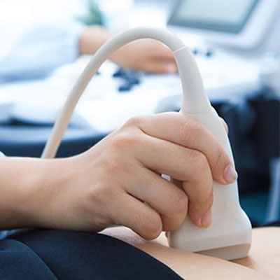
How is an ultrasound is performed?
The Doctor (Radiologist) usually holds a transducer, a hand-held device, like a wand, which is placed on the patient’s skin. A water based gel is applied to the skin which helps transmit the sound waves. Depending on the area being examined, you may need to change positions. After the procedure is over, the gel will be cleaned off of your skin.
Ultrasound Scans performed at Kataria Healthcare
- Upper Abdomen
- Lower abdomen/ Pelvis
- Transvaginal scan
- Whole Abdomen
- KUB
- Transrectal Ultrasound for Prostate(TRUS)
- Follicular study
- Obstetric Scan
- Early Pregnancy
- Level I or NB / NT scan – This can be combined with double marker
- Level II or Anomaly Scan
- Interval Growth Scan
- BPP Scan/Manning Score
- Fetal Doppler/Obs Colour Doppler
- High frequency Scans – Breast, Thyroid, Scrotum/Testis, Joints, hernia etc.
- Ultrasound guided FNACs
- Venous colour doppler for Upper and Lower limbs
- Arterial colour doppler for Upper and Lower limbs
- Renal doppler
- Carotid doppler
- 3D/4D/5D Ultrasound: Experience the miracle of watching your unborn baby in 4D/5D HD LIVE realistic views.
- Shear and strain wave Elastography.
- Liver Elastography: Liver fibrosis quantification.
- Liver UGAP score: Liver fat quantification.