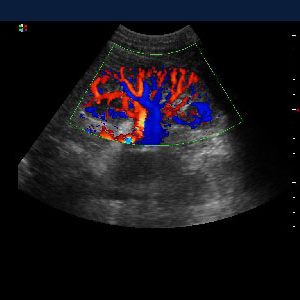
Colour Doppler is a special type of ultrasound which uses the computer analytics to convert measurements into an array of colours to visualize the speed and direction of blood flow through a vessel.
It is a risk-free and pain-free procedure. The transducer sends sound waves through your skin and other body tissues to the blood vessels. The sound waves echo off of your blood vessels and send the information to a computer to be processed and recorded. The computer will produce graphs or pictures that show the flow of the blood through your arteries and veins.
Why is a Doppler Ultrasound performed?
The Doppler ultrasound exam may be ordered if you show signs of:
- A blood clot forming in a vein deep inside your body resulting in pain and swelling.
- Varicose veins and incompetent valves of the blood vessels.
- Narrowing and hardening of the arteries that supply blood to the legs and feet
- Vascular tumours/swellings.
- To look for clots in neck vessels supplying the brain.
- Diagnose any vascular compromise in kidneys in patients with hypertension.
- In pregnancy to know about maternal and fetal circulation and predict growth restriction.
What happens during a Doppler Ultrasound?
- You will need to remove clothing, jewellery, and any other objects in the area that will be studied and may be asked to wear a hospital gown.
- You’ll be instructed about the position depending on the part to be assessed.
- A water-soluble gel will be put on a transducer, which directs high-frequency sound waves into the arteries or veins being studied.
- Images are taken as the transducer is moved along the part being studied. You may hear a “whooshing” sound as blood flow is detected.
Dopplers at Kataria Healthcare
- Carotid
- Renal
- Scrotum
- Arterial-upper/lower limbs
- Venous-upper/lower limbs
- Abdominal
- Uterus
- Pregnancy/obs/fetal